
Fractures in Animals: Types, Signs, Healing Stages & Treatment (Complete Vet Guide)
1. INTRODUCTION. Successful treatment of fracture requires better understanding of anatomy & physio-pathologic reactions, which occur in the injured tissues. The science & art of bone & joints surgery requires an understanding of mechanical forces involved in operative surgery. The strength of bone & amount of external violence exerted govern occurrence and type of fracture. Fracture is manifested by some predominant signs such as pain, palpable breakage, crepitating sound on mobilization and the hemorrhage in advance cases. Shortly after the fracture, the blood fills the fracture site & surrounding with haematoma, which later on coagulates. Primary callus is formed & the formations of bony trabeculae (cancellous bone) directly by osteoblast & indirectly by replacement of cartilage by bone, result in bone union. The callus between the fragment is reabsorbed & replaced by compact bone. Process of reconstruction of the bone is very slow & may take several months or up to year. There is tremendous body response to the fracture. Enzyme secretion & calcium mobilization to the fracture site occurs in the initial phase. Diagnosis of clinical union is manifested by absence of sign of pain & immobility at the fractured site. Confirmatory diagnosis is always made by radiography. Some important complications occur during the healing process, the most significant of which is delayed healing & non-union of fracture. In order to observe the successful treatment procedure,s all the possible causes leading to delayed healing or non-union must be addressed soon after the fracture has occurred.
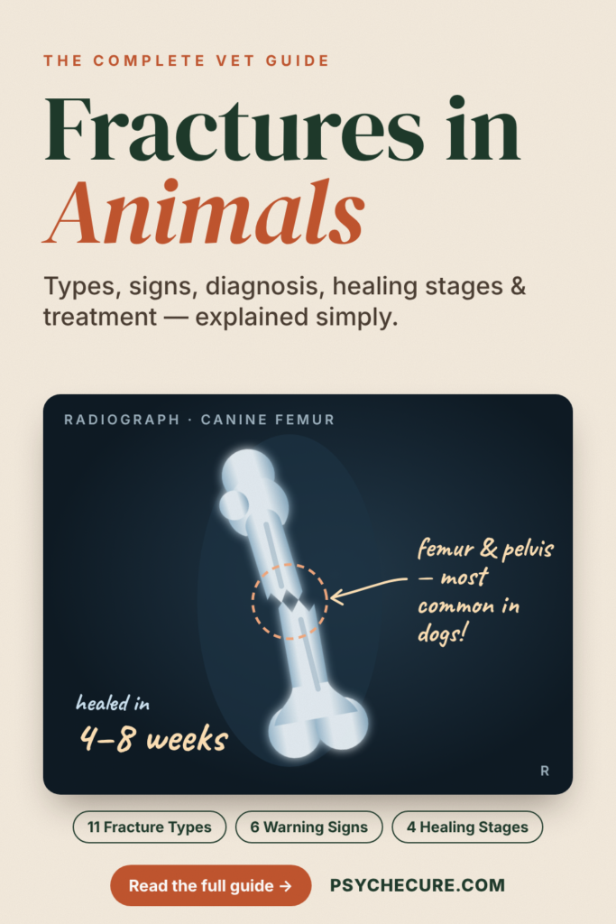
2. FRACTURE. A Fracture is the break in the continuity of a bone and it may be complete or incomplete disruption in the continuity of a bone. Strength of normal bone is related to the age of the animal. Occurrence & type of fracture is governed by strength of a bone, the amount and, type of exertion & point of trauma causing fracture.
3. ETIOLOGY
- Due to anatomical position of bones & their vulnerability to the fracture like their superficial position & shape.
- External causes like trauma, fall, kicks etc.
- Diseased conditions of bone like osteoporosis, & ostitis.
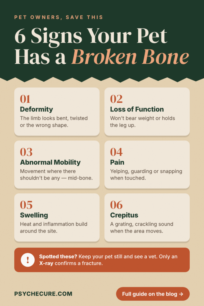
4. SIGNS OF FRACTURE. Following are signs of fracture: –
a. Deformity
b. Loss of function
c. Abnormal mobility
d. Pain
- Inflammation
- Crepitating sound on mobilization
5. DIAGNOSIS. Diagnosis is made from:-
a. Signs of fracture.
b. Radiography.
6. CLASSIFICATION OF FRACTURE. Fractures are broadly classified into three classes, namely simple, compound & complicated fracture.
a. Simple Fracture. A fracture, which does not communicate with the out side. There is no wound on skin leading to the fracture site. It is also called closed fracture.
- Incomplete Fracture. A fracture, which does not extend through complete thickness of the bone. e.g. Green stick fractures, fissured fracture & deferred fracture.
(2) Green Stick Fracture. A fracture in which the bone is partly broken.
(3) Fissured Fracture. There are fissures or cracks through bone without causing displacement.
b. Compound Fracture (Open Fracture). A fracture, which is communicated with the out side with open wound on the skin, is called compound fracture. A fracture in which the wound is caused at the site of fracture either from external trauma or by the protrusion of bone fragments through the skin. When a simple fracture has little been rendered compound, it is called secondary compound fracture.
c. Complicated Fracture. A closed fracture in which there is a considerable injury to the important neighboring vessels or nerve is accompanied by opening of a joint or visceral cavity is called as complicated fracture, e.g. complicated fracture of ribs.
d. Complete Fracture. Fractures can also be classified as;
(1) Simple Fracture. When the bone is broken in one place.
(2) Double Fracture. When there are two fractures in one bone.
(3) Multiple Fractures. Where the bone is broken into more than two pieces.
(4) Transverse Fracture. A fracture at right angle to the axis of the bone.
(5) Longitudinal Fracture. A fracture extending in the longitudinal direction.
(6) Colles Fracture. Fracture of distal end of the radius.
(7) Pathological Fracture. A fracture of bone occurring due to some disease & not by trauma.
(8) Spiral Fracture. It is found in spiral direction.
(9) Oblique Fracture. A fracture extending in an oblique direction.
(10) Impacted Fracture. A fracture in which one fragment is firmly driven into the fracture site of another fracture e.g. head of femur being driven into acetabulum.
- Dentate Fracture. A fracture in which ends of the fragments are toothed & interlocked.
7. PATHOLOGY OF A FRACTURE & BODY RESPONSE. If the reparative processes involved in fracture repair are to be appreciated fully, the pathology of a fracture must be visualized. The following body responses occur in the body to a fracture and the secondary soft tissue trauma at the cellular level.
- a. Hyperemia. It occurs shortly after the fracture of bone.
- b. Haematoma. It is formed at the site of fracture.
c. Deposition of Calcium. Calcium is mobilized from bone, without changing the blood calcium level. This process is continued until hyperemia subsides.
- Enzyme Secretion. The enzyme phosphatase is secreted by proliferating cartilage cells and osteoblasts reaching max 6-8 times higher of haematoma. This excess persists throughout the active growth of cartilage cells and osteoblasts.
- Acidic pH. During the 1st or 2nd week of the fracture the pH of haematoma remains acidic but later on becomes alkaline for the deposition of calcium. There is no appreciable change in blood calcium level.
- Alkaline pH. Acidic pH turns into alkaline pH at which the deposition of calcium takes place and the primary bony callus is formed.
- Calcification. It takes place on the 10th day & the fracture healing becomes visible radiographically.
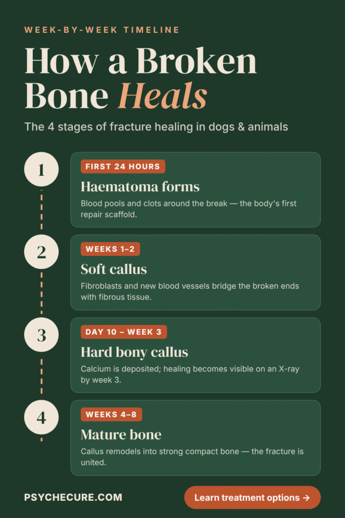
- HEALING OF FRACTURE. In dogs the most common fracture is the fractures of femur & pelvis. Healing of fracture is described in four stages: –
a. Haematoma Formation. Fracture causes injuries to the blood vessels & surrounding soft tissues causing hemorrhages. This blood collect around the sides of fracture forming a haematoma later on which coagulates. The haematoma is well-formed with-in 24 hours.
b. Callus Formation. Within 24 hour the growth of fibroblast & capillaries into clot starts. The growth of capillaries takes place from periosteum, haversion canals end-osteum and bone marrow. Macrophages (wandering phagocytic cells) make their way into blood clot to remove the excessive RBC & wasted debris. The clot at this stage contains fibrin, fibroblast & newly formed blood vessels & resembles granulation tissues. This fibro vascular tissue thus formed between the broken ends of fracture is called soft callus or fibrous callus or temporary callus. The callus fills the place between the broken ends. The size of the callus is large when the hemorrhage at the fracture site is extensive. The soft callus is formed with in one or two weeks. The vascularity of granulation tissues is diminished when it is converted into fibrous callus.
c. Primary Bone Callus Formation. During the early stages of the fracture of bone, reaction of clot is acidic. The presence of cellular debris consisting of damaged tissue and hemorrhage is responsible for lowering the pH. The acidity favor the mobilization of calcium from bone fragments as calcium phosphorus and from the blood. The enzyme “phosphatase” released by the osteoclasts also facilitates release of calcium phosphate from plasma so as to cause the supersaturation of calcium in the haematoma surrounding the fractured site. Even though there is no appreciable variation in blood calcium level. The calcium mobilized from the bone fragments and plasma is held in the solution and the pH remains acidic. Calcium is later on deposited when the reaction slowly turns alkaline. It results in the formation of primary bone callus. Osteoblast derived from the bone fragments invades the callus and calcium is deposited in the intercellular space. Osteoclasts are the more numerous in periosteum and endo-steum and extremely few in the compact bone. Therefore mineralization takes place more at the ends (Peripheral) & central zone of the callus while it is meager at the intermediary zone. The mineralization at the peripheral & middle zone affords a temporary union & immobilizes the fragments until long tissue is formed later. The primary callus now prevents a granular appearance and feels like an irregular hard mass of cartilage & bone. Calcification commences on the 10th day of fracture & about the 3rd week it becomes visible radiographically. Clinically it can be felt as firm & round mass around the site of fracture. With in 4-8 weeks depending on the size of fracture, bone and age of patient, the primary callus becomes sufficiently firm to prevent any movement. It is clinically called “The union of fractured bone”. No pair is evident on movement at this stage.
d. Mature Bone Formation. It takes place from 4-8 weeks.
9. FACTORS AFFECTING HEALING. Following factors effects the healing of fracture: –
- Size of fracture.
- Size of bone.
- Age of the patient.
- Alkaline reaction essential for deposition of calcium.
- Site of fracture.
- Removal of all devitalized tissues.
- Reduction (Restoration of proper alignment).
- Fixation (immobilization).
j. Rest.
10. DIAGNOSIS OF CLINICAL HEALING. Clinical healing is that stage of healing of fracture when the fixation device is removed. It is manifested by:
a. Palpable hard irregular callus.
b. Absence of signs of pain, when force is applied.
c. Immobility at fracture site.
d. In case of questionable physical examination, the radiographic examination is applied.
- As immature bone cells have relatively low mineral contents they are not readily seen on the radiographic study. Demonstrable radiographic appearance of bony tubercle across the fracture confirms the presence of union & assures that remodeling has taken place. Over- lapping fragments or impacted fragments may stimulate bony union.
- Formation of primary callus takes place with in 48 hrs. Cellular proliferation begins in the periosteum, & haversian canal.
- Fragments of the tissue degenerate & disintegrate & removed by phagocytes.
j. The periosteum undergoes thickening & the dead bone near the fracture site undergoes demineralization & re-absorption.
- Re-absorption, replacement & remodeling & callus bone connecting the bone fragments is gradually replaced by mature bone.
9.
11. REPAIR AND REDUCTION METHODS. The treatment of any fracture and dislocation involves the application of certain orthopedic principles as much as it does that of some internal and external device for the immobilization of the fracture or reduction of a dislocation.
a. The correction of displacement of fractured end of the bone so as to bring the fragments into normal alignment and apposition is called “Reduction of Fracture.” If the displacement is slight, holding the upper segments of bone & then moving the lower segments into correct position can do reduction.
b. In case of marked displacement as in over riding fracture, it is done under general anesthesia. The over riding can be usually corrected by extension & counter extension combined with local manipulation. Exerting traction on the distal fragment is the extension or traction. Traction should be exerted in the direction of limb.
c. Counter extension is the procedure adopted for pulling the proximal fragments in the opposite direction. This can be affected by fixing the upper part of the limb while the lower part of the limb is being pulled.
d. Two methods of reduction are generally employed to treat a fracture, namely, closed and open reduction.
(1) Closed reduction. Reduction is accomplished with the integument intact. Exceptions to this definition exist when halfpin splintage, treatment of compound fractures by closed reduction, and closed intramedullary pinning are employed. The technique employed to achieve closed reduction may involve the use of angulations or toggling of fragments during manipulation to reduce the fracture. Immobilization of the reduced fracture is achieved by use of the following equipment:
- Gordon extender
(b) Thomas splint
(c) Yucca-board splint
(d) Plaster-of-Paris bandage
(e) Metasplint
(f) Orthoclase splint
- Half pin splintage
- Compression bar splintage
- Closed intramedullary pinning
(2) Closed reduction is suitable only if the bone fragments can be palpated in order to be aligned. Excessive soft-tissue swelling prohibits the effective use of closed reduction; if it is attempted, further serious soft-tissue damage may result. The wise clinician thinks little of offering sedation to the patient to permit time for swelling to subside before the reduction and immobilization is attempted.
(3) Open Reduction. Open reduction involves incising the integument to expose the fracture site in order to achieve reduction. Angulations and toggling may still have to be employed to reduce the fracture, but the likelihood of causing unseen soft-tissue damage is lessened with open reduction. This method also allows the surgeon to manipulate the fragments with hand tools and levers. Once open reduction has been accomplished, immobilization can be maintained with a variety of internal and/or external appliances. All the equipment listed to immobilize fractures treated by closed reduction can be utilized in the open-reduction technique, as well as the following:
- Smillie nails
- Lag screw
- Cancellous screw
- Wire sutures
- K wire
- Intramedullary pins
- Rush pins
- Transfixion screws combined with halfpin splintage, full pin splintage, compression plates or a compression bar
(j) Half pin or full pin splintage
(k) Neutralization plate
(l) Compression plate
(m) Tension-band wire
e. Retention & Immobilization. It includes fixing the bone fragments so that they are motionless with respect to each other during healing process. The object is to fix the fragments in desired position & prevent displacement, angulation and rotation. Various aids as already described are used like POP. Intramedullary pin & bone plates.
12. DELAYED & NON-UNION OF FRACTURE. Complication during healing which influence healing may occur at the site of fracture in the adjacent soft tissue or in the tissue & organs at the distance. Complications at the fracture site include delayed union or non-union, angular deformity or malunion, shortening of the bone & alteration in growth, vascular necrosis & infection. Delayed union & non-union is summarized as under:
a. Delayed Union. It is associated with limited osteogenic activity & healing process is prolonged. Radiographic features of delayed union are: –
(1) The fracture line is wide & clearly evident.
(2) External & Internal calluses are minimal.
(3) The bone at the fracture site has feathery or wooly appearance.
b. Non Union. It is manifested to the condition when all the osteogenic activities at the fracture site have been diminished & union is no longer possible. Radiographic feature of non-union is: –
- Rounding of the bone ends with sclerosis at the fracture site.
- A well defined gap between the fragments.
- Gradual bowing at the fracture site particularly if the weight bearing has been allowed.
- Demineralization of bone above & below the fracture site, particularly if the bone is not in use.
- Closure of modularly canals in both the fragments.
c. Clinical Signs of Delayed & Non Union. The clinical signs associated with Non union are:-
(1). Lack of rigidity or bowing at the fracture site.
(2). Excessive soft tissue callus or minimal callus.
(3). Evidence of pain when the bone is used.
- d. Types of Fracture Susceptible to Delayed or Non Union
(1). Transverse or short oblique fractures of distal half of radius & ulna.
(2). Fracture of femoral neck.
(3). Fracture of the distal 3rd of tibia.
(4). Transverse mid shaft fracture.
e. Causes of Delayed or Non Union
- (1). Inadequate reduction.
(2). Inadequate immobilization or failure to maintain it until union is complete.
(3). Impairment of blood supply due to trauma or damage inflicted by surgeon.
(4). Infection.
(5). Distraction of fragments.
(6). Loss of bone from open fracture (at the time of surgery).
(7). Motion or weight bearing permitted too early.
f. Treatment of Delayed/Non Union
(1). Evaluation of method of fixation clinically, If delayed.
(2). Reduction & maintenance of reduction for absolute immobility or for an extended period is sufficient to promote union.
(3). Stimulation of osteogensis if still delayed.
(4). In case of unsatisfactory reduction, it should be improved after exposing the fracture site.
(5). Stimulation of osteogensis by bone grafting. Various grafts (inlay, diamond inlay, massive onlay, onlay, intra medulary peg) may be applied.
(6). Fresh bone may be obtained from the patient, or osteogenous graft from the donor of the same species. A rib or the crest of the ilium is easily accessible source, bone may be collected & held for future use. It should be obtained aseptically placed in sterile tube & sealed. It is then placed in the freezer at –5 to –20 F such bone may be used for years long.
(7). Surgical Treatment: if the position of the fracture is satisfactory but the union is delayed, then the broken end may be perforated with many drill holes to aid revascularization. It should be followed by rigid uninterrupted fixation.
(8). If the fragments are immobilized the graft require a min fixation. The graft should be rigidly affixed, however, if the fragments are not accompanying firm fixation, then additional fixation should be applied externally.
13. OTHER COMPLICATIONS DURING HEALING
- Avascular Necrosis of Bone. In avascular necrosis the bone dies because its blood supply is reduced. This occurs mostly when the fracture is close to articular surface or after a distribution, which has disrupted blood supply. Mostly occur in small dogs. It may be associated with osteomyelitis. Cystic appearance of the bone distal to epiphysial line in case of aseptic femur necrosis & some time cystic appearance with distortion manifest it.
- Mal-union & Angular Deformity. These deformities are usually the results of faulty reduction & fixation but some time unavoidable. When crushing injuries involving the end of a bone, then the patient is kept under observation for several months. In case of deformity of proximal or distal portion of bone, functions are severely impaired. Angular deformity may result from injuries or abnormal growth near the epiphysial line of radius & ulna in growing dog resulting in the out word deviation of & rotation of the foot. The defect usually becomes pronounced. The animal by favouring the leg places more pressure on the medial than the lateral side, which retard growth on the lateral surface of the radius & accelerates on the medial side.
- Shortening of the Bone. Shortening of a long bone occurs in many fractures, but the animal accommodates it by increased extension of joints of the limb. Shortening is more serious when it occurs at the distal end of radius & ulna. Causes of shortening are: –
(1). The fragment units with overriding.
(2). The fragment of a bone is lost.
(3). Fragments are compressed at fracture site.
(4). Injuries to an epiphysial plate in growing animal result in premature closure.
- Vascular Injury. Major blood vessels get injured when the fracture occurs or at the time of treatment. Clinical signs include pallor, coldness, swelling & diminished pulse. End to end anastomosis of vessel is feasible. Intact vessels may undergo spastic contraction resulting in stenosis. Histologically appearance is same as that of muscle infarction or ischemia with varying degree of necrosis.
- Nerve Injury. Three types of nerve injuries occur:
(1). Complete division of nerve resulting in paralysis.
(2). Damage to axon without disturbing the continuity of nerve, peripheral degeneration follows & recovery is slow.
(3). Simple contusion with temporary loss of nerve function resulting recovery with in week or two.
f. Joints Stiffness. Joint stiffness associated with fracture occurs due to capsular adhesion, which results from accumulation of exudates in particular tissues.
14. CONCLUSION. Bone commonly fractured in dogs are femur & pelvis mostly due to trauma or external violence. As soon as the fracture of bone takes place, corrective measures must be taken to enhance the natural healing process. There is spontaneous healing of fracture after its occurrence distributed at various time intervals, formation of haematoma & with in 48 hours of the fracture primary callus formation. It is followed by formation of soft callus. There is differentiation of & proliferation of osteogenic cells & proliferation of haversion canal, periosteum & endosteum. Phagocytosis of degenerative tissue occurs & the periosteum undergoes thickening. It is then followed by formation of primary bone callus and pain is still evident on movement. Formation of secondary bone callus & function of mature bone is finally seen with in or up to 4-8 weeks. Ossification takes place with in this period. In order to adhere to proper treatment of wound fracture, external causes leading to various complications of healing must be removed. Of course the use of some aid & regular checking & treatment will end in sound healing process.
By Dr Muhammad Usman Bashir